- In-Stock Tumor Cell Lines
- Human Orbital Fibroblasts
- Human Microglia
- Human Pulmonary Alveolar Epithelial Cells
- Human Colonic Fibroblasts
- Human Type II Alveolar Epithelial Cells
- Human Valvular Interstitial Cells
- Human Thyroid Epithelial Cells
- C57BL/6 Mouse Dermal Fibroblasts
- Human Alveolar Macrophages
- Human Dermal Fibroblasts, Adult
- Human Lung Fibroblasts, Adult
- Human Retinal Muller Cells
- Human Articular Chondrocytes
- Human Retinal Pigment Epithelial Cells
- Human Pancreatic Islets of Langerhans Cells
- Human Kidney Podocyte Cells
- Human Renal Proximal Tubule Cells
Colon-related diseases are a kind of diseases with high incidence rate and serious impact on people’s safety and quality of life. Therefore, colon diseases are a prominent research field in clinical medicine and basic medicine.
Introduction: Colon and Colonic epithelium
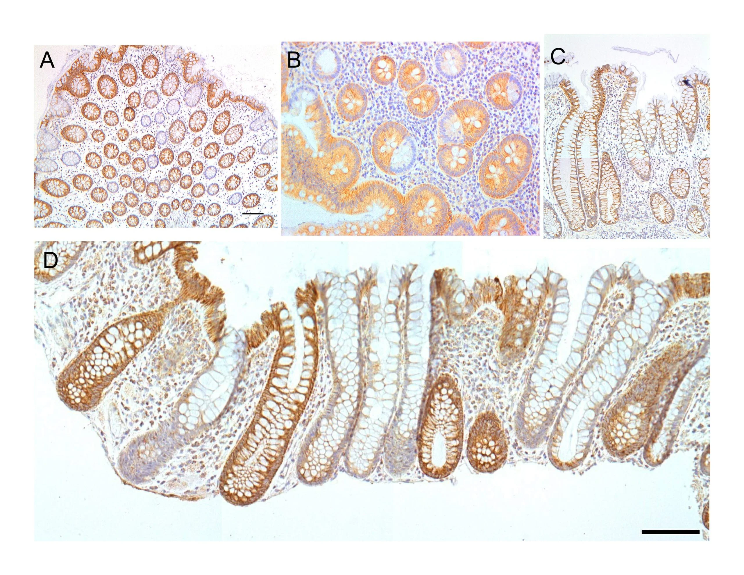
The colon is the longest part of the large intestine, which is part of the last segment of the digestive tract and the last part of the digestive system[1]. (Figure.1) The functions of the colon mainly include the final extraction of water and residual nutrients from digested residual waste and serving as a site for the colonization and fermentation of the gut microbiome[2]. Colonic epithelium serves as a mechanical and immunology barrier of the intestinal wall, shows the functions of coordinates digestion and absorption, and provides a venue for gut microbiome colonization[3; 4]. The colonic epithelium is mainly composed of the simple columnar epithelium (columnar epithelial cells) and invaginations called crypts. Stem cells that maintain the regenerative ability of the epithelium are distributed at the bottom of the crypt, while differentiated and mature colonic epithelial cells gradually migrate upwards from the bottom of the crypt until shedding in the colon cavity[5]. (Figure.2) In addition to columnar epithelial cells, there are also a large number of goblet cells in the colon epithelium, which play an important role in mucosal maintenance and immune regulation[6].

Figure.1 Large intestine and colon[7].

Figure.2 Colonic epithelial crypt[8].
Colonic epithelium is closely related to the main type of colon disease, inflammatory bowel disease (IBD). IBD is the general name for a series of intestinal diseases, mainly including colitis and Crohn’s disease (CD). The main cause is the disruption of homeostasis between gut microbiota and epithelial mucosal immunity[3]. Various factors such as infection, disruption of gut microbiota, autoimmune factors, diet, abnormal bile acids metabolism, and mechanical damage to the epithelial mucosa may lead to the occurrence of IBD[9]. IBD is a precursor disease to colorectal cancer (CRC). CRC is a kind of malignant disease, predominantly related to aging and poor lifestyle habits (which can also act as a trigger for IBD), while genetic factors do not account for the majority[10]. The five-year survival rate of CRC is 60%, indicating that CRC is a disease with high mortality and difficult to treat. Active prevention and early detection are the most effective ways to reduce mortality[11].
In Vitro Model of Colonic Epithelium and Human Colonic Epithelial Cells
Optional in vitro models in the research of colon physiology and pathology mainly include immortalized cell lines, tumor cell lines, primary cells, and organoids. Immortalized cell lines and tumor cell lines are the most widely used due to their efficient cultivation and straightforward application, but long-term generation and immortalization can lead to deviations in the genetic background compared to in vivo models, thereby affecting the reliability of data. Therefore, primary cells, especially human primary cells, are frequently found in high-impact publications to enhance data credibility. Furthermore, organoid culture based on primary colon stem cells is an advanced in vitro model that has emerged in recent years. It can partially restore the structure of colon epithelium in vitro when the genetic background is close to the body, providing robust support for a detailed analysis of physiological or pathological processes[12]. However, although organoids are a recently developed technology, they are not widely applied due to their high cost and technical difficulties. In contrast, primary cells are frequently used models in research work.
In this section, we briefly introduce the isolation, extraction, and culture protocols of human primary colonic epithelial cells[12; 13]. Firstly, the mucosa was stripped from clinical biopsy tissue and washed several times with PBS. If stem cells are needed, the biopsy tissue needs a complete crypt structure. After cleaning the mucus, the mucosa was placed in Hank’s balanced salt solution with 2mM EDTA but without calcium and magnesium, and rotated three times for 10 min at 37°C. The supernatant after the second and third rotations was retained. The primary colonic epithelial cells were precipitated from the supernatant, and washed several times with PBS, and then cultured with medium in 37℃, 5% CO2. The culture medium contains 10% or 20% FBS and growth hormone (such as EGF), and the specific formula can be explored based on the requirements of cell growth.
Application of Human Colonic Epithelial Cells in the Publications
Human colonic epithelial cells is frequently used to validate crucial data obtained from immortalized or tumor cell lines to demonstrate the reliability of the data. In recent high-impact publications, in vitro organoid culture based on human primary colon epithelial cells has become increasingly employed. Parikh et al. analyzed the colonic epithelial cells’ diversity in health and IBD through primary human colonic epithelial cells[3]. The researchers isolated the cells from clinical biopsy, and performed single-cell sequencing and flow cytometry analysis. By combining the result from animal models and mouse-derived organoid models, they identified the fundamental determinants of epithelial barrier failure in IBD. In another study, Dutton et al. identified that TNF-α but not hyperglycemia is the main cause of colon epithelial dysfunction[14]. The researchers established a human colon epithelial in vitro model based on human primary colonic epithelial cells and hydrogel support, and achieved a more precise mechanism analysis of intestinal epithelial dysfunction.
Conclusion
Colon-related diseases plague the lives and quality of life of countless patients, and malignant diseases can lead to fatalities. Thus, colon diseases and physiological mechanisms are one of the hotspots in medical research. Human primary epithelial cells show immense value in the research, they can provide reliable data and are the foundation for emerging organoid in vitro culture techniques, enabling it worthwhile for researchers to master their application methods.
Where to Get Digestive System Primary Cells for Your Research?
AcceGen isolates and offers a wide range of high-quality digestive system primary cells, such as Human Colonic Epithelial Cells, Human Intestinal Epithelial Cells, Human Small Intestine Epithelial Cells, and Human Large Intestine Epithelial Cells. These cell products provide you with a convenient means to research. To get more information, please refer to: Digestive System Primary Cells.
It is our pleasure to help relative researches to move forward. All the products of AcceGen are strictly comply with international standards. For more detailed information, please visit our product portfolio or contact inquiry@accegen.com.
References
[1] L.L. Azzouz, and S. Sharma, Physiology, Large Intestine, StatPearls, StatPearls Publishing
Copyright © 2023, StatPearls Publishing LLC., Treasure Island (FL), 2023.
[2] D. Krogh, Biology: A Guide to the Natural World, Benjamin Cummings, 2011.
[3] K. Parikh, A. Antanaviciute, D. Fawkner-Corbett, M. Jagielowicz, A. Aulicino, C. Lagerholm, S. Davis, J. Kinchen, H.H. Chen, N.K. Alham, N. Ashley, E. Johnson, P. Hublitz, L. Bao, J. Lukomska, R.S. Andev, E. Björklund, B.M. Kessler, R. Fischer, R. Goldin, H. Koohy, and A. Simmons, Colonic epithelial cell diversity in health and inflammatory bowel disease. Nature 567 (2019) 49-55.
[4] L.W. Peterson, and D. Artis, Intestinal epithelial cells: regulators of barrier function and immune homeostasis. Nat Rev Immunol 14 (2014) 141-53.
[5] A.M. Baker, B. Cereser, S. Melton, A.G. Fletcher, M. Rodriguez-Justo, P.J. Tadrous, A. Humphries, G. Elia, S.A. McDonald, N.A. Wright, B.D. Simons, M. Jansen, and T.A. Graham, Quantification of crypt and stem cell evolution in the normal and neoplastic human colon. Cell Rep 8 (2014) 940-7.
[6] J.R. McDole, L.W. Wheeler, K.G. McDonald, B. Wang, V. Konjufca, K.A. Knoop, R.D. Newberry, and M.J. Miller, Goblet cells deliver luminal antigen to CD103+ dendritic cells in the small intestine. Nature 483 (2012) 345-9.
[7] https://en.wikipedia.org/wiki/Large_intestine#Structure.
[8] 
[9] D.C. Baumgart, and S.R. Carding, Inflammatory bowel disease: cause and immunobiology. Lancet 369 (2007) 1627-40.
[10] A.J. Watson, and P.D. Collins, Colon cancer: a civilization disorder. Dig Dis 29 (2011) 222-8.
[11] D. Cunningham, W. Atkin, H.J. Lenz, H.T. Lynch, B. Minsky, B. Nordlinger, and N. Starling, Colorectal cancer. Lancet 375 (2010) 1030-47.
[12] O. Dmitrieva-Posocco, A.C. Wong, P. Lundgren, A.M. Golos, H.C. Descamps, L. Dohnalová, Z. Cramer, Y. Tian, B. Yueh, O. Eskiocak, G. Egervari, Y. Lan, J. Liu, J. Fan, J. Kim, B. Madhu, K.M. Schneider, S. Khoziainova, N. Andreeva, Q. Wang, N. Li, E.E. Furth, W. Bailis, J.R. Kelsen, K.E. Hamilton, K.H. Kaestner, S.L. Berger, J.A. Epstein, R. Jain, M. Li, S. Beyaz, C.J. Lengner, B.W. Katona, S.I. Grivennikov, C.A. Thaiss, and M. Levy, β-Hydroxybutyrate suppresses colorectal cancer. Nature 605 (2022) 160-165.
[13] K. Schlottmann, F.P. Wachs, J. Grossmann, D. Vogl, M. Maendel, W. Falk, J. Schölmerich, T. Andus, and G. Rogler, Interferon gamma downregulates IL-8 production in primary human colonic epithelial cells without induction of apoptosis. International Journal of Colorectal Disease 19 (2004) 421-429.
[14] J.S. Dutton, S.S. Hinman, R. Kim, P.J. Attayek, M. Maurer, C.S. Sims, and N.L. Allbritton, Hyperglycemia minimally alters primary self-renewing human colonic epithelial cells while TNFα-promotes severe intestinal epithelial dysfunction. Integr Biol (Camb) 13 (2021) 139-152.

Copyright - Unless otherwise stated all contents of this website are AcceGen™ All Rights Reserved – Full details of the use of materials on this site please refer to AcceGen Editorial Policy – Guest Posts are welcome, by submitting a guest post to AcceGen you are agree to the AcceGen Guest Post Agreement – Any concerns please contact marketing@accegen.com


