MONO-MAC-6
Discover top-quality products tailored for scientific and medical research. Request a personalized quote today
to enhance your projects.
MONO-MAC-6 is a human monocyte cell line derived from patients with acute monocytic leukemia. MONO-MAC-6 cells possess phenotypic and functional features of mature monocytes such as CD14 expression, phagocytic capacity, and cytokine production. Acute monocytic leukemia (AMoL or AML-M5) is a type of acute myeloid leukemia characterized by peripheral monocytosis. More than 80% of the nucleated cells of AMoL are of monocytic origin. Studies demonstrate that MONO-MAC-6 cells exhibit similar monocyte-endothelial interaction to human monocytes.
Why choose MONO-MAC-6 from AcceGen?
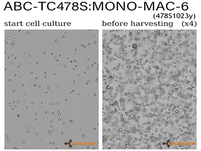
MONO-MAC-6 cells are established from the peripheral blood of a 64-year-old man with relapsed acute monocytic leukemia. Cells are cryopreserved to ensure the best viability. Each vial contains more than 1×10^6 cells in 1ml volume. MONO-MAC-6 cells carry a t(9;11)(p22;q23) translocation, which results in the fusion of KMT2A-MLLT3 gene (MLL-MLLT3; MLL-AF9). Subculture cells at a split ratio of 1:3 to 1:5 with a cell seeding density between 0.3×10^6 to 1×10^6 cells/ml.
Product Code | MONO-MAC-6; Mono-mac-6; MONO-MAC 6; Mono Mac 6; Mono Mac6; MonoMac 6; MonoMac6; MONOMAC6; MM6 |
Species | Human |
Cat.No | ABC-TC478S |
Quality Control | All cell lots test negative for HIV-1, HBV, HCV, mycoplasma, yeast, fungi, and other pathogens. |
Product Category | Tumor Cell Lines |
Size/Quantity | 1 vial |
Shipping Info | Dry Ice |
Growth Conditions | 37 ℃, 5% CO2 |
Disease | Acute Monocytic Leukemia |
Storage | Liquid Nitrogen |
Product Type | Human Leukemia Cell Lines |
When you publish your research, please cite our product as “AcceGen Biotech Cat.# XXX-0000”. In return, we’ll give you a $100 coupon. Simply click here and submit your paper’s PubMed ID (PMID).
For research use only
MONO-MAC-6 is a useful cell model for in vitro studies of monocyte biology and physiology. They can also be used for cytogenetic analysis of gene translocation, gene expression profiling, drug screening assay and testing response towards various molecules.
Gentle Handling: Use a serological pipette to gently resuspend cells. Avoid vigorous pipetting to prevent mechanical stress.
Passage Protocol: Collect cells by transferring the culture medium (containing suspended cells) to a centrifuge tube. Centrifuge at 300×g for 5-10 minutes. Aspirate the supernatant carefully and resuspend the cell pellet in fresh pre-warmed complete medium. Seed the cells at the desired density into new culture flasks.
Macrophage Markers: CD11b, CD14, CD68, and F4/80.
Dendritic Cell Markers: CD11c, HLA-DR, CD80, CD83, and CD86.
Flow Cytometry: Use flow cytometry to quantify the expression of these surface markers.
RT-PCR and Western Blot: Assess gene and protein expression levels of differentiation markers.
Issue 1: Poor Differentiation
Solution: Ensure the correct concentration and timing of differentiation factors. Validate the activity of the growth factors or supplements.
Issue 2: Cell Death
Solution: Optimize the concentration of differentiation agents and minimize handling stress. Ensure proper medium composition and culture conditions.
Issue 3: Heterogeneous Differentiation
Solution: Sort differentiated cells using flow cytometry to obtain a more homogeneous population.