Home > Products > Tumor Cell Lines > Human Tumor Cell Lines >
MDA-MB-361 | ||||
---|---|---|---|---|
Product Name | MDA-MB-361 | |||
Price | Get Quote | |||
Product Code | MDA-MB 361; MDA MB 361; MDA-MB361; MDAMB361; MDA-361; MDA361; MB361; MD Anderson-Metastatic Breast-361 | |||
Cat.No | ABC-TC0652 | Species | Human | |
Size/Quantity | 1 vial | Biosafety Level | 1 | |
Shipping Info | Dry Ice | Storage | Liquid Nitrogen | |
Description | This line differs from others of the series in karyology and in that it was isolated from a brain metastasis. | |||
Source Organ | Breast | |||
Recommended Medium And Supplement | L-15 Medium + 10% FBS | |||
Citation Guide | When you publish your research, please cite our product as “AcceGen Biotech Cat.# XXX-0000”. In return, we’ll give you a $100 coupon. Simply click here and submit your paper’s PubMed ID (PMID). | |||
Application | For research use only | |||
Growth Conditions | 37 ℃ | |||
Cell Type | Epithelial | |||
Growth Mode | Adherent | |||
Product Type | Human Breast Cancer Cell Lines | |||
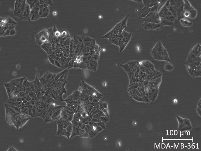
Product Image |
Frequently Asked Questions
What are MDA-MB-361 cells?
MDA-MB-361 cells were originally isolated from metastatic site in the brain of a patient with breast adenocarcinoma. Cells are transplantable to mouse hypodermis but not to abdominal.
Is MDA-MB-361 cell line triple-negative?
The MDA-MB-361 cells are characterized as ER-positive/Progesterone receptor (PgR) negative, luminal mammary carcinoma.
How do I passage MDA-MB-361 cells?
Procedure
Preparation:
Warm the L15 medium, PBS, and trypsin-EDTA solution to 37°C in a water bath.
Ensure all necessary equipment and reagents are sterile and ready for use.Observe Cells:
Place the flask containing ells under an inverted microscope.
Check for cell confluence (typically, cells should be 70-80% confluent before passaging).Remove Old Medium:
In a biosafety cabinet, aspirate the old medium from the flask carefully without disturbing the cell monolayer.Rinse Cells:
Add 5-10 mL of PBS to the flask to rinse off any residual medium and serum that may inhibit trypsin activity.
Gently rock the flask to ensure the cells are thoroughly rinsed, then aspirate the PBS.Add Trypsin-EDTA:
Add enough trypsin-EDTA solution to cover the cell monolayer (typically 1-2 mL for a T-75 flask).
Gently tilt the flask to ensure the entire surface is covered with trypsin-EDTA.Incubate:
Place the flask in the incubator for 3-5 minutes, checking the cells under the microscope periodically. The cells should round up and detach from the surface.Neutralize Trypsin:
Once the cells are detached, add 5-10 mL of complete L15 medium (with FBS) to the flask to neutralize the trypsin.Collect Cells:
Gently pipette the cell suspension up and down to ensure all cells are detached and in suspension.
Transfer the cell suspension to a 15 mL conical tube.Centrifuge:
Centrifuge the cell suspension at 200 x g for 5 minutes to pellet the cells.Resuspend Cells:
Carefully aspirate the supernatant without disturbing the cell pellet.
Resuspend the cells in an appropriate volume of fresh complete medium.Count Cells (Optional):
If needed, take a small aliquot of the cell suspension to count cells using a hemocytometer or an automated cell counter.Seed New Flasks:
Seed the cells into new culture flasks or plates at the desired density. For example, if splitting at a 1:5 ratio, add 1 part of the cell suspension to 4 parts of fresh medium in a new flask.Incubate:
Place the newly seeded flasks or plates back into the incubator set at 37°C without CO2 (or use non-vented flask and keep the lid closed).Monitoring:
Observe the cells under the microscope after 24 hours to ensure they have adhered and are spreading out. Change the medium every 2-3 days or as needed.What is the karyotype of MDA-MB-361 cell line?
The model number of MDA-MB-361 cell line is 56, with a range of 54-61.
- ONLINE INQUIRY
- PRODUCT REVIEWS
Fill out a request in the form below and we’ll get back to you within 24 hours with a quote.