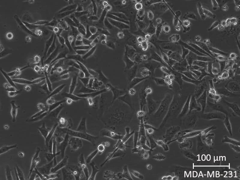
1. Culture Medium
Base Medium: Use Dulbecco’s Modified Eagle Medium (DMEM) or RPMI-1640. Both media are commonly used for MDA-MB-231 cells.
Serum: Supplement the medium with 10% Fetal Bovine Serum (FBS). Ensure the FBS is heat-inactivated to remove complement proteins that could affect cell growth.
Antibiotics: Add 1% Penicillin-Streptomycin to prevent bacterial contamination.
2. Culture Conditions
Temperature: Maintain the cells at 37°C in a humidified atmosphere with 5% CO2.
pH: Ensure the pH of the culture medium is around 7.2-7.4, as this is optimal for MDA-MB-231 cell growth.
Osmolality: Keep the osmolality of the culture medium between 280-320 mOsm/kg.
3. Seeding Density
Initial Seeding: Seed cells at a density of 2.5-5 × 10^4 cells/cm². Adjust density depending on the growth rate and the specific needs of your experiments.
Subculturing: When cells reach 70-80% confluence, they should be subcultured to prevent overgrowth and maintain healthy proliferation.
4. Subculturing Protocol
Remove Old Medium: Aspirate the old culture medium carefully.
Rinse with PBS: Wash the cells with PBS without calcium and magnesium to remove residual serum that can inhibit trypsin activity.
Trypsinization: Add enough trypsin-EDTA solution (0.25%) to cover the cell monolayer and incubate for 2-5 minutes at 37°C until the cells detach.
Neutralize Trypsin: Add complete medium (with FBS) to neutralize the trypsin and collect the cells.
Centrifuge: Spin down the cells at 200×g for 5 minutes.
Resuspend and Seed: Resuspend the cell pellet in fresh medium and seed into new flasks at the appropriate density.
5. Monitoring and Maintenance
Medium Changes: Replace the culture medium every 2-3 days to ensure cells have adequate nutrients and to remove waste products.
Morphology Check: Regularly observe cells under a microscope to check for healthy morphology and detect any signs of contamination or differentiation.
Cell Counting: Periodically count the cells using a hemocytometer or an automated cell counter to monitor growth rates and adjust seeding densities as needed.
6. Supplementation (if needed)
Growth Factors: In specific experiments, adding growth factors like Epidermal Growth Factor (EGF) or Insulin-like Growth Factor 1 (IGF-1) can stimulate proliferation and signal transduction studies. Typically, 10-20ng/mL for EGF and 10-50ng/mL for IGF-1.
Glutamine: Ensure the medium is supplemented with L-glutamine (2mM) as it is essential for cell growth and viability.
7. Special Considerations
Thawing and Cryopreservation: Thaw cells quickly in a 37°C water bath and handle them gently. For freezing, use a cryoprotectant solution such as 90% FBS and 10% DMSO, and freeze cells slowly using a controlled-rate freezing container.
Authentication: Regularly authenticate cell lines using STR profiling to ensure the identity of the cells and check for mycoplasma contamination.


