1
Discover top-quality products tailored for scientific and medical research. Request a personalized quote today
to enhance your projects.
Product Code | HRA |
Species | Human |
Cat.No | ABC-TC3787 |
Quality Control | All cells test negative for mycoplasma, bacteria, yeast, and fungi. |
Product Category | Primary Cells |
Size/Quantity | 1 vial |
Cell Type | Astrocyte |
Shipping Info | Dry Ice |
Growth Conditions | 37 ℃, 5% CO2 |
Source Organ | Retina |
Disease | Normal |
Biosafety Level | 1 |
Storage | Liquid Nitrogen |
Product Type | Ocular Cells |
Retinal astrocytes are a type of glial cells that are located in the retinal nerve fiber and ganglion cell layers and provides them with intrinsic protection and support. In human retina, there are two types of astrocytes can be distinguished, that is elongated astrocytes and star-shaped astrocytes. Both astrocytes are variants or subclasses of a single type of astrocyte, specializing in different ways. Star-shaped astrocytes specialize more in relations with the blood vessels and elongated astrocytes with the axons. The recent studies show that transcription factor Pax8 can be as a highly specific marker of retinal astrocytes.
Why choose Human Retinal Astrocytes from AcceGen?
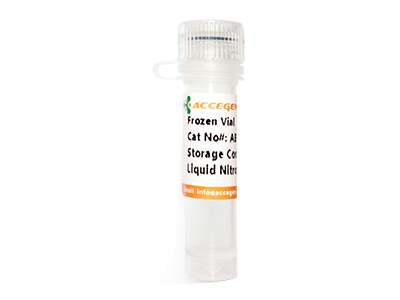
Human Retinal Astrocytes (HRA) from AcceGen are isolated from human retina and cryopreserved at early passage. HRA are cryopreserved in a cryogenic vial, providing a convenient storage format and are guaranteed to further expand for 15 population doublings under the conditions provided. The cell density per tube is more than 5×10⁵/ml. HRA are negative for mycoplasma.
When you publish your research, please cite our product as “AcceGen Biotech Cat.# XXX-0000”. In return, we’ll give you a $100 coupon. Simply click here and submit your paper’s PubMed ID (PMID).
FOR THE RESEARCH USE ONLY
Retinal astrocytes are responsible for the maintenance of the retinal microenvironment and structural support. They are involved in retinal glucose metabolism, providing retinal neurons with the necessary nutrients for oxidative metabolism. They are also involved in regulating proper immune responses. In retinopathy, astrocytes adopt defensive programs, which is disease- and context-specific and involves the activation of thousands of genes to maintain retinal homeostasis.
Studies have shown that astrocytes can trigger the inflammation in glaucoma. Under glaucoma, they become reactive and change their phenotypes into neurotoxic ones. They release neurotoxic factors such as cytokines and chemokines and damage axons of retinal ganglion cells.
Human Retinal Astrocytes are glial cells found in the retina, where they provide structural and functional support to neurons and blood vessels. They play a key role in maintaining retinal homeostasis, regulating the blood-retinal barrier, and responding to injury or disease.
When the retina is damaged, retinal astrocytes become reactive, proliferating and releasing signaling molecules that mediate inflammation and repair. This process, known as gliosis, can either support regeneration or contribute to scarring depending on the extent of injury.
Yes, Human Retinal Astrocytes are valuable in modeling retinal diseases, particularly those involving neurodegeneration and vascular dysfunction. They are used to study conditions such as glaucoma, age-related macular degeneration, and retinal ischemia.
These cells are cultured in a specialized medium enriched with factors that support astrocyte growth and function, such as neurotrophic factors. They thrive in environments at 37°C with 5% CO2, mimicking the physiological conditions of the retina.
Retinal astrocytes play a key role in neurovascular coupling, which is the process by which neuronal activity regulates blood flow in the retina. They mediate communication between neurons and blood vessels, ensuring adequate blood supply in response to retinal activity.