1
Discover top-quality products tailored for scientific and medical research. Request a personalized quote today
to enhance your projects.
Product Code | HCPEpiC |
Species | Human |
Cat.No | ABC-TC3568 |
Quality Control | All cells test negative for mycoplasma, bacteria, yeast, and fungi. |
Product Category | Primary Cells |
Size/Quantity | 1 vial |
Cell Type | Epithelial |
Shipping Info | Dry Ice |
Growth Conditions | 37 ℃, 5% CO2 |
Source Organ | Choroid Plexus |
Disease | Normal |
Biosafety Level | 1 |
Storage | Liquid Nitrogen |
Product Type | Nervous Cells |
Key Features | -Backed by AcceGen advanced technology |
In central nervous system, the choroid plexus acts as a barrier that separates the blood from cerebrospinal fluid (CSF). It is composed of cuboidal epithelium surrounding capillaries that embedded in a loose connective tissue. The choroid plexus epithelial cells (CPECs), linked by tight junctions, form a neuroprotective barrier by regulating the exchange of electrocytes, nutrients, metabolic wastes, and xenobiotics into and out of the central nervous system. The choroid plexus epithelium is responsible for synthesis and secretion of CSF, regulation of hormone signaling and passage of leukocytes during brain injury and infections. Morphological changes such as atrophy of choroidal epithelial cells, are commonly seen in aging and neurological disorders such as Alzheimer disease.
Why choose Human Choroid Plexus Epithelial Cells from AcceGen?
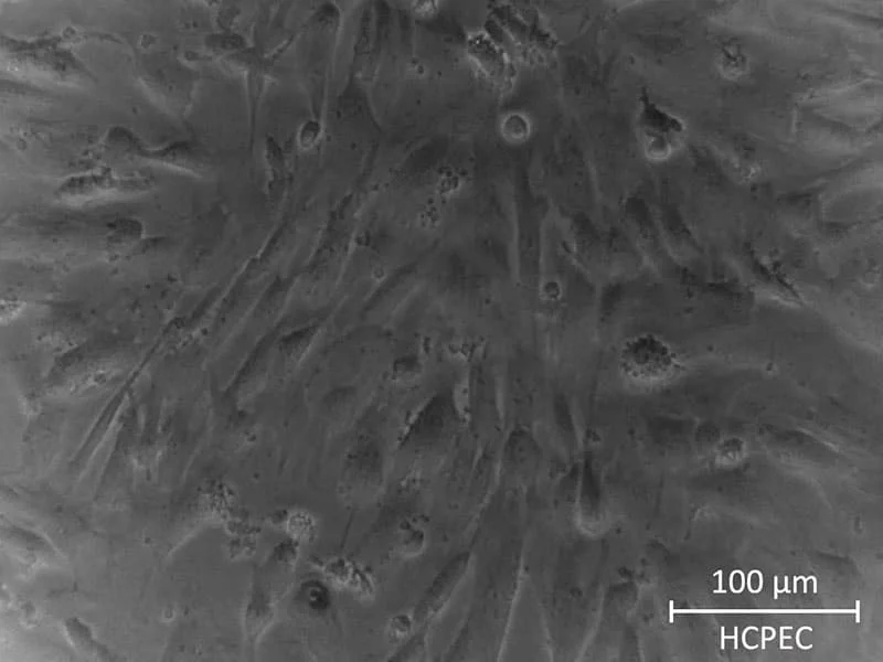
Human choroid plexus epithelial cells are derived from human choroid plexus. Cells are cryopreserved immediately after purification and delivered in dry ice. Each vial contains > 5×10^5 cells in 1 ml volume. Choroidal epithelial cells are characterized with antibodies specific to cytokeratin-18, -19 and vimentin. Cells are tested negative for HIV-1, HBV, HCV and other pathogens. Human choroid plexus epithelial cells are quality tested and guaranteed to further expand for 5 population doublings when cultured in AcceGen recommended cell culture media.
When you publish your research, please cite our product as “AcceGen Biotech Cat.# XXX-0000”. In return, we’ll give you a $100 coupon. Simply click here and submit your paper’s PubMed ID (PMID).
Erb, U., Schwerk, C., Schroten, H., & Karremann, M. (2020). Review of functional in vitro models of the blood-cerebrospinal fluid barrier in leukaemia research. Journal Of Neuroscience Methods, 329, 108478. https://doi.org/10.1016/j.jneumeth.2019.108478
For research use only
Human Choroid Plexus Epithelial Cells are specialized cells found in the choroid plexus, a structure within the brain’s ventricles responsible for producing cerebrospinal fluid (CSF). These cells form a barrier between the blood and CSF, and regulate the composition of CSF by transporting ions and nutrients.
The main function of these cells is to produce and regulate the composition of cerebrospinal fluid (CSF). They maintain the blood-CSF barrier, transport ions, and remove waste products from the brain, playing a vital role in maintaining the brain’s homeostasis.
These cells form tight junctions that create the blood-CSF barrier, which prevents harmful substances in the blood from entering the CSF. They selectively allow the transport of ions, nutrients, and other molecules into the CSF while blocking toxins and pathogens.
These cells are used in research related to cerebrospinal fluid production, blood-CSF barrier integrity, neuroinflammation, and brain homeostasis. They are also utilized in drug discovery to develop therapies for diseases that affect the choroid plexus or CSF regulation.
Yes, these cells are valuable for modeling neurological diseases, particularly those involving disruptions in CSF production or blood-CSF barrier dysfunction. They are used to study neuroinflammatory diseases, hydrocephalus, and central nervous system infections.