Discover top-quality products tailored for scientific and medical research. Request a personalized quote today
to enhance your projects.
Product Code | BV2 |
Species | Mouse-C57BL/6 |
Cat.No | ABC-TC212S |
Product Category | Tumor Cell Lines |
Size/Quantity | 1 vial |
Cell Type | Microglial |
Shipping Info | Dry Ice |
Growth Conditions | 37 ℃, 5% CO2 |
Source Organ | Brain |
Storage | Liquid Nitrogen |
Product Type | Mouse Brain Cancer Cell Lines |
Key Features | -Backed by AcceGen advanced technology |
BV-2 Cells Derivation and biomarker
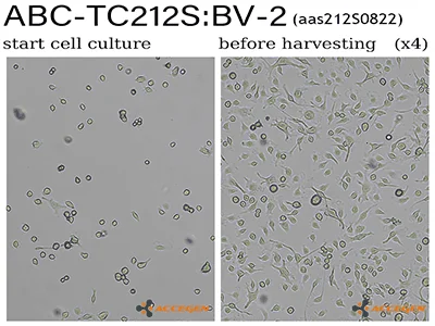
BV-2 is a type of microglial cell derived from C57/BL6 murine. The BV2 cells are immortalized by v-raf/v-myc carrying J2 retrovirus. BV2 express nuclear v-myc and the cytoplasmic v-raf oncogene products as well as the env gp70 antigen at the surface level. BV2 microglia cell line retains microglia morphological and functional characteristics. In accordance with v-raf/v-myc expressed characters, the metabolic and proliferation rate of in vitro BV-2 greatly exceeds that of other microglia.
Why Choose BV-2 from AcceGen?
AcceGen BV-2 cells are positive for MAC1 and MAC2 antigens, negative for MAC3, glial fibrillary acidic protein (GFAP) and galactocerebroside (GC).
When you publish your research, please cite our product as “AcceGen Biotech Cat.# XXX-0000”. In return, we’ll give you a $100 coupon. Simply click here and submit your paper’s PubMed ID (PMID).
Hagan, N., Kane, J.L., Grover, D., Woodworth, L., Madore, C., Saleh, J., Sancho, J., Liu, J., Li, Y., Proto, J. and Zelic, M., 2020. CSF1R signaling is a regulator of pathogenesis in progressive MS. Cell death & disease, 11(10), pp.1-25.
Lee, H.J., Seo, M., Baek, M., Shin, Y.P., Lee, J.H., Kim, I.W., Hwang, J.S. and Kim, M., 2020. Inhibitory Effect of Protaetiamycine 6 on Neuroinflammation in LPS-stimulated BV-2 Microglia. Journal of Life Science, 30(12), pp.1078-1084.
Inada, M., Xu, H., Takeuchi, M., Ito, M. and Chen, M., 2021. Microglia increase tight-junction permeability in coordination with Müller cells under hypoxic condition in an in vitro model of inner blood-retinal barrier. Experimental Eye Research, 205, p.108490.
Liu, Y., Hammel, G., Shi, M., Cheng, Z., Zivkovic, S., Wang, X., Xu, P., He, X., Guo, B., Ren, Y. and Zuo, L., 2021. Myelin debris stimulates NG2/CSPG4 expression in bone marrow-derived macrophages in the injured spinal cord. Frontiers in Cellular Neuroscience, 15, p.80.
Li, J., Cheng, X., Fu, D., Liang, Y., Chen, C., Deng, W. and He, L., 2022. Autophagy of Spinal Microglia Affects the Activation of Microglia through the PI3K/AKT/mTOR Signaling Pathway. Neuroscience, 482, pp.77-86.
2022). Imaging inflammasome activation in microglia. Current Protocols, 2, e578. doi: 10.1002/cpz1.578.
Lee, J.H., Ji, S.H., Lim, J.S. et al. Anti-neuroinflammatory Effects and Brain Pharmacokinetic Properties of Selonsertib, an Apoptosis signal-regulating Kinase 1 Inhibitor, in mice. Neurochem Res 47, 3829–3837 (2022). https://doi.org/10.1007/s11064-022-03777-9.
Hasriadi et al. “Antinociceptive efficacy of Clerodendrum petasites S. Moore, a Thai medicinal plant, and its CNS safety profiles.” Journal of traditional and complementary medicine vol. 13,1 81-92. 12 Nov. 2022, doi:10.1016/j.jtcme.2022.11.001.
Lim, Jae Sung, et al. “In Vitro Anti-Inflammatory Effects of Symplocos Sumuntia Buch.-Ham. Ex D. Don Extract via Blockage of the NF-ΚB/JNK Signaling Pathways in LPS-Activated Microglial Cells.” Plants, vol. 11, no. 22, Nov. 2022, p. 3095. Crossref, https://doi.org/10.3390/plants11223095.
Janpaijit, Sakawrat et al. “Anti-neuroinflammatory effects of Cleistocalyx nervosum var. paniala berry-seed extract in BV-2 microglial cells via inhibition of MAPKs/NF-κB signaling pathway.” Heliyon vol. 8,11 e11869. 28 Nov. 2022, doi:10.1016/j.heliyon.2022.e11869.
For research use only
BV-2 cells are a murine microglial cell line commonly used as a model for studying microglial function and neuroinflammation. They offer a consistent and reproducible system for investigating the immune responses of microglia in the central nervous system.
Medium: Use high-glucose DMEM supplemented with 10% fetal bovine serum (FBS), L-glutamine, and antibiotics (penicillin-streptomycin).
Incubation: Maintain cells at 37°C in a humidified atmosphere with 5% CO₂.
Medium Renewal: Change the medium every 2-3 days to ensure optimal nutrient availability and waste removal.
Lipopolysaccharide (LPS): Often used to activate microglial cells and induce an inflammatory response.
Interferon-gamma (IFN-γ): Enhances the activation and pro-inflammatory response of microglia.
Tumor Necrosis Factor-alpha (TNF-α): Used to study inflammatory signaling pathways.