1
Discover top-quality products tailored for scientific and medical research. Request a personalized quote today
to enhance your projects.
Product Code | B16-BL6; B16 BL6; B16BL6; B16BL-6; BL6; BL-6; B16-F10-BL6 |
Species | Mouse |
Cat.No | ABC-TC0060 |
Product Category | Tumor Cell Lines |
Size/Quantity | 1 vial |
Shipping Info | Dry Ice |
Growth Conditions | 37 ℃, 5% CO2 |
Source Organ | Skin |
Disease | Melanoma |
Biosafety Level | 1 |
Storage | Liquid Nitrogen |
Product Type | Mouse Melanoma Cell Lines |
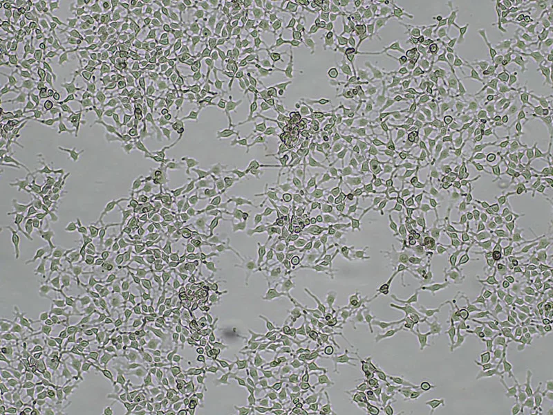
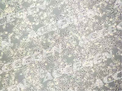
The B16-BL6 cell line is a murine melanoma model derived from B16-F10, known for its tissue-invasive property. Clones derived from B16-BL6 exhibit high metastatic potential and elevated levels of hormonally-stimulated adenylate cyclase, confirming the connection between hormonal responsiveness and metastasis. These melanoma cells are poorly immunogenic, similar to other spontaneously arising tumors, and express low levels of major histocompatibility complex (MHC) antigens. Additionally, B16-BL6 cells release inhibitory factor(s) that affect the active pump activity in isolated lymph vessels.
Why choose B16-BL6 from AcceGen?
B16-BL6 from AcceGen is the ideal choice due to its sterile nature and ability to incubate under standardized conditions. With high quality and viability, it boasts an advanced cryopreservation technique ensuring optimal preservation. Additionally, AcceGen employs rigorous quality control measures, further enhancing the reliability and consistency of the B16-BL6 cell line.
When you publish your research, please cite our product as “AcceGen Biotech Cat.# XXX-0000”. In return, we’ll give you a $100 coupon. Simply click here and submit your paper’s PubMed ID (PMID).
Clement, E., Lazar, I., Attané, C., Carrié, L., Dauvillier, S., & Ducoux‐Petit, M. et al. (2020). Adipocyte extracellular vesicles carry enzymes and fatty acids that stimulate mitochondrial metabolism and remodeling in tumor cells. The EMBO Journal, 39(3). https://doi.org/10.15252/embj.2019102525
FOR RESEARCH USE ONLY
The B16-BL6 cell line serves as a valuable tool for studying various aspects of melanoma, including metastasis, hormonal regulation, and tumor immune evasion. Additionally, this cell line finds application in assessing the efficacy and pharmacodynamics (PD) of anti-cancer therapeutics. Researchers can utilize the B16-BL6 model in both subcutaneous and metastatic settings, allowing for the investigation of tumor growth, response to treatments, and the development of novel therapeutic strategies. Its availability in different models enhances its versatility and utility in preclinical studies focused on melanoma research and drug development.
The C57BL/6 melanoma cell line B16/BL6 was derived from the B16-F10 model for its more tissue-invasive property. What distinguishes the B16/BL6 cell line from its parent B16-F10 cell line is its ability to cross the bladder membrane.
The B16/BL6 cell line is pivotal for melanoma modeling progression and new therapies testing. Its applications include evaluating the effectiveness of drug candidates, understanding metastatic behavior, and studying immune responses.
Procedure
Preparation:
Warm the RPMI 1640 medium, PBS, and trypsin-EDTA solution to 37°C in a water bath.
Ensure all necessary equipment and reagents are sterile and ready for use.
Observe Cells:
Place the flask containing MDA-MB-453 cells under an inverted microscope.
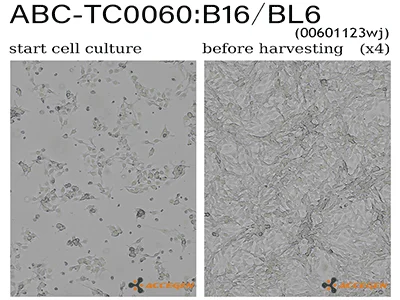
Check for cell confluence (typically, cells should be 70-80% confluent before passaging).
Remove Old Medium:
In a biosafety cabinet, aspirate the old medium from the flask carefully without disturbing the cell monolayer.
Rinse Cells:
Add 5-10 mL of PBS to the flask to rinse off any residual medium and serum that may inhibit trypsin activity.
Gently rock the flask to ensure the cells are thoroughly rinsed, then aspirate the PBS.
Add Trypsin-EDTA:
Add enough trypsin-EDTA solution to cover the cell monolayer (typically 1-2 mL for a T-75 flask).
Gently tilt the flask to ensure the entire surface is covered with trypsin-EDTA.
Incubate:
Place the flask in the incubator for 3-5 minutes, checking the cells under the microscope periodically. The cells should round up and detach from the surface.
Neutralize Trypsin:
Once the cells are detached, add 5-10 mL of complete RPMI 1640 medium (with FBS) to the flask to neutralize the trypsin.
Collect Cells:
Gently pipette the cell suspension up and down to ensure all cells are detached and in suspension.
Transfer the cell suspension to a 15 mL conical tube.
Centrifuge:
Centrifuge the cell suspension at 200×g for 5 minutes to pellet the cells.
Resuspend Cells:
Carefully aspirate the supernatant without disturbing the cell pellet.
Resuspend the cells in an appropriate volume of fresh complete RPMI 1640 medium.
Count Cells (Optional):
If needed, take a small aliquot of the cell suspension to count cells using a hemocytometer or an automated cell counter.
Seed New Flasks:
Seed the cells into new culture flasks or plates at the desired density. For example, if splitting at a 1:5 ratio, add 1 part of the cell suspension to 4 parts of fresh medium in a new flask.
Incubate:
Place the newly seeded flasks or plates back into the incubator set at 37°C with 5% CO2.
Monitoring:
Observe the cells under the microscope after 24 hours to ensure they have adhered and are spreading out. Change the medium every 2-3 days or as needed.